

Case Library
Explore Patient Assessment Cases
Browse through a wide range of patient cases with comprehensive assessments, evaluation scales, and clinical findings.


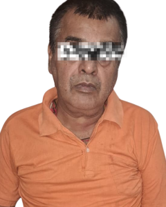
Right pca stroke
About condition
Medical Diagnosis
Subacute right posterior cerebral artery (PCA) territory ischemic infarction involving the right parieto-occipito-temporal region, with bilateral thalamic and right midbrain involvement, associated with complete right PCA occlusion/non-opacification, likely thrombotic in etiology.
patient details
“A 56-year-old right-hand-dominant male shopkeeper, 3–4 months post right PCA territory ischemic stroke, with left hemiparesis, hypertension and Type 2 diabetes mellitus, presenting for neurological physiotherapy rehabilitation.”
Chief Complaints
Patient presents with:
- Difficulty in walking.
- Weakness of the left upper and lower limb.
- Dragging of the left foot during walking.
- Left foot drop.
- Difficulty in bed mobility.
- Difficulty during sit-to-stand and other transfers.
- Mild left shoulder subluxation.
- Knee pain, particularly around the affected knee.
- Pulling/stretching sensation around the groin/hip–knee region during movement.
- Difficulty performing some ADLs independently.
History of Present Illness
The patient is a 56-year-old male, known case of hypertension and recently diagnosed Type 2 Diabetes Mellitus.
According to the available history, the patient developed sudden onset of dizziness around 25–26 March. Following the episode of dizziness, he developed sudden weakness initially in the left lower limb, which subsequently progressed to involve the left upper and lower limbs.
Initially, the patient experienced temporary improvement after receiving local medication. However, after approximately two days, he again developed dizziness with worsening of weakness. He was subsequently referred for further neurological evaluation.
CT scan and CT angiography revealed an ischemic infarction in the right posterior cerebral artery (PCA) territory with complete non-opacification of the right PCA, suggestive of occlusion/thrombosis.
The patient was admitted to the ICU for approximately one week. Following stabilization, he was shifted to the ward and later discharged.
At present, approximately 3–4 months after the stroke, the patient is conscious and cooperative but continues to have left-sided weakness, foot drop, impaired gait, reduced lower-limb control and difficulty with functional mobility.
Medical History
Past Medical History
- Hypertension – present.
- Type 2 Diabetes Mellitus – diagnosed around the time of stroke.
- Right PCA ischemic stroke – present.
- ICU admission – approximately one week.
No significant history of:
- Previous head trauma
- Major surgery
- Previous neurological disease
- Previous seizure disorder
- Loss of consciousness
Medication History
Patient is reportedly taking Levera (levetiracetam) as prescribed.
Medication details should be verified from the patient's current prescription/medical record rather than assumed from history.
Personal & Occupational History
| Parameter | Finding |
|---|---|
| Occupation | Shopkeeper |
| Diet | Regular diet |
| Appetite | Normal |
| Sleep | No major disturbance reported |
| Bowel | No incontinence reported |
| Bladder | No incontinence reported |
| Pre-morbid activity | Shop-related/community activities |
| Current occupational status | Limited due to mobility impairment |
Pain Assessment
Site
- Left knee region
- Pulling sensation around groin/hip–knee region
Nature
- Dull aching pain
- Pulling/stretching sensation
Onset
Pain developed during the post-stroke recovery period.
Aggravating factors
- Walking
- Lower-limb movement
- Weight-bearing
- Functional activities
Relieving factors
- Rest
- Reduction in activity
Duration
Intermittent.
Severity
NPRS: 4 during sle
Important clinical point
Because the pain is around the medial/superior patellar region, the examination should differentiate among:
- Quadriceps tendon involvement
- Patellofemoral pain
- Medial patellar soft-tissue involvement
- Knee OA
- Muscle tightness/abnormal loading secondary to hemiparetic gait
General Observation
Build
Average build.
Consciousness
Patient is conscious and cooperative.
Orientation
Oriented to:
- Person
- Place
- Time
Posture
Abnormal postural alignment secondary to left-sided weakness.
Attitude
Protective/guarded use of affected side due to weakness.
External signs
- Left hemiparesis
- Mild shoulder subluxation
- Left foot drop
- Reduced voluntary movement
- Abnormal gait
Involuntary movements
Not observed.
Trophic changes
No significant trophic changes noted.
Skin
Appears intact.
Edema
No significant edema reported/observed.
Systemic Examination
A. Respiratory System
- Breathing pattern: Thoraco-abdominal
- Chest symmetry: Present
- Chest expansion: Bilaterally symmetrical
- Respiratory distress: Absent
B. Cardiovascular System
- History of hypertension present.
- Pulse regular.
- Peripheral edema observed.
- BP monitoring required.
C. Musculoskeletal System
Problems identified:
- Left-sided weakness
- Shoulder subluxation
- Knee pain
- Foot drop
- Reduced ankle dorsiflexion
- Muscle tightness
- Reduced functional mobility
D. Gastrointestinal System
- Appetite normal.
- No bowel incontinence reported.
- Regular oral diet.
E. Genitourinary/Autonomic System
- No bladder dysfunction reported.
- No bowel dysfunction reported.
neurological examination
Level of Consciousness
| Component | Finding | Score |
|---|---|---|
| Eye opening | Spontaneous | 4/4 |
| Verbal response | Oriented/converses appropriately | 5/5 |
| Motor response | Obeys commands | 6/6 |
| Total | Conscious and oriented | 15/15 |
Interpretation
GCS = 15/15, indicating the patient is conscious and appropriately responsive.
Cognitive Assessment
| Function | Finding |
|---|---|
| Orientation | Intact |
| Short-term memory | Intact |
| Long-term memory | Intact |
| Attention | Adequate |
| Judgment | Appropriate |
| Calculation | Able to perform simple calculations |
| Following commands | Intact |
| Communication | Understandable |
Cranial Nerve Examination
| Cranial Nerve | Examination | Finding |
|---|---|---|
| CN I | Smell | Intact/reported intact |
| CN II | Vision/visual field | No disturbance reported |
| CN III, IV, VI | Eye movements | Intact |
| CN V | Facial sensation/mastication | Intact |
| CN VII | Facial movement | Finding should be objectively reassessed |
| CN VIII | Hearing/balance | No major abnormality reported |
| CN IX, X | Swallowing/voice | Intact |
| CN XI | Shoulder shrug | May be reduced on affected side |
| CN XII | Tongue movement | Intact |
Important
Because this is a right PCA stroke, visual-field assessment is particularly important even if the patient does not spontaneously report visual problems.
Recommended examination:
- Confrontation visual-field testing
- Visual scanning
- Functional visual assessment
Sensory Examination
| Modality | Finding |
|---|---|
| Light touch | Intact / mildly decreased – to confirm |
| Pain | Intact |
| Temperature | Intact |
Deep Sensation
| Modality | Finding |
|---|---|
| Joint position | Intact |
| Vibration | Intact |
| Kinesthesia | Intact |
Cortical Sensation
| Test | Finding |
|---|---|
| Two-point discrimination | Intact |
| Stereognosis | Intact |
| Graphesthesia | Intact |
| Tactile localization | Intact |
Interpretation
No definite major sensory deficit is documented based on the available findings.
Motor Examination
Muscle Tone
Initial stage
Patient was initially described as flaccid following stroke.
Current stage
There is a possible transition toward increased tone.
Modified Ashworth Scale
Grade 0–1 – mild increase in tone may be present.
However, individual muscle groups should be scored separately rather than assigning one MAS grade to the whole limb.
Muscle Power Examination – MMT
| Muscle Group | Right | Left |
|---|---|---|
| Shoulder flexion | 5/5 | 3+/5 |
| Shoulder abduction | 5/5 | 3+/5 |
| Elbow flexion | 5/5 | 4−/5 |
| Elbow extension | 5/5 | 4−/5 |
| Wrist movement | 5/5 | 3+/5 |
| Hip flexion | 5/5 | 3+/5 |
| Knee extension | 5/5 | 4−/5 |
| Ankle dorsiflexion | 5/5 | 2–3/5 |
| Ankle plantarflexion | 5/5 | 3+/5 |
Interpretation
Patient demonstrates left-sided motor weakness, with more significant distal weakness at the ankle, particularly dorsiflexion.
The ankle dorsiflexion weakness is clinically consistent with the observed left foot drop.
Voluntary Motor Control
The patient demonstrates:
- Reduced selective motor control on the left.
- Difficulty isolating movements.
- Reduced distal motor control.
- Emerging voluntary movement.
- Difficulty controlling the affected lower limb during gait.
Brunnstrom Recovery
Based on the available description, the patient appears to be in an intermediate recovery phase with voluntary movement emerging, but a definite Brunnstrom stage should be assigned only after testing synergy, isolated movement and movement out of synergy systematically.
| Reflex | Right | Left |
|---|---|---|
| Biceps | +2 | +2 |
| Triceps | +2 | +2 |
| Supinator | +2 | +2 |
| Knee jerk | +2 | +3 |
| Ankle jerk | +2 | +3 |
| Plantar | Flexor | Extensor tendency – to confirm |
Interpretation
Brisk left knee and ankle reflexes may indicate upper motor neuron involvement/evolving hyperreflexia following stroke.
Plantar response should be formally reassessed and documented.
Coordination Assessment
Non-equilibrium
- Finger-to-nose: Mild difficulty on affected side.
- Finger opposition: Slow/impaired.
- Heel-to-shin: Difficult due to weakness.
- Rapid alternating movements: Reduced speed.
Equilibrium
- Romberg: Mild instability.
- Tandem standing: Difficult.
- Single-leg standing: Unable on affected side.
- Functional reach: Reduced.
Range of Motion
| Joint | Finding |
|---|---|
| Shoulder | Mild restriction associated with subluxation |
| Elbow | Functional ROM |
| Wrist | Functional ROM |
| Fingers | Functional ROM |
| Joint | Finding |
|---|---|
| Hip | Mild restriction due to pain/tightness |
| Knee | Mild painful ROM |
| Ankle | Reduced dorsiflexion |
| Muscle | Finding |
|---|---|
| Hamstrings | Mild tightness |
| Gastrocnemius–soleus | Tightness |
| Hip flexors | Mild tightness |
| Pectoralis muscles | Possible tightness |
Balance Assessment
Static Balance
Sitting: Good
Standing: Fair
Single-leg stance: Impaired on left.
Dynamic Balance
Patient demonstrates difficulty with:
- Turning
- Gait initiation
- Weight shifting
- Maintaining stability during walking
Gait Assessment
Type
Hemiparetic gait
Major deviations
- Left foot drop
- Foot dragging during swing phase
- Reduced left stance time
- Reduced step length
- Reduced weight acceptance on affected limb
- Compensatory hip/knee movement
- Difficulty with gait initiation
- Possible circumduction/hip hiking due to foot clearance problem
Main contributing factors
- Left dorsiflexor weakness
- Reduced selective motor control
- Possible increased plantar-flexor tone
- Reduced balance
- Reduced weight-bearing confidence
- Hip/knee weakness
- Muscle tightness
Gait Assessment
Bed Mobility
Patient has difficulty with:
- Rolling
- Supine-to-sitting
- Repositioning
- Moving the affected lower limb
Requires assistance/supervision.
Transfers
Sit-to-stand
Possible but difficult.
Problems may include:
- Reduced left weight bearing
- Quadriceps weakness
- Poor balance
- Reduced motor control
Bed-to-chair
Requires supervision/assistance.
Activities of Daily Living
| Activity | Functional Status |
|---|---|
| Feeding | Independent |
| Grooming | Independent/minimal assistance |
| Dressing | Difficulty |
| Bathing | Assistance required |
| Toileting | Supervision |
| Walking | Difficult |
| Stairs | Difficult |
| Community mobility | Restricted |
| Shop-related activities | Restricted |
ICF-Based Physiotherapy Diagnosis
Body Structure & Function
Body structures involved:
- Brain – right PCA territory
- Left upper limb
- Left lower limb
- Shoulder complex
- Knee/ankle
Impairments:
- Muscle weakness
- Abnormal tone
- Foot drop
- Pain
- Reduced ROM
- Impaired balance
- Reduced coordination
- Reduced motor control
Activity
Patient has difficulty with:
- Walking
- Transfers
- Bed mobility
- Dressing
- Bathing
- Stair negotiation
Participation
Patient has difficulty:
- Returning to occupation
- Community mobility
- Social activities
- Independent daily life
Environmental factors
Potential facilitators:
- Family assistance
- Physiotherapy
- AFO
- Walking aid
- Home modification